
E Ample Of Normal Chest Ray Report
E Ample Of Normal Chest Ray Report - Regularly reviewing and presenting radiographs to senior colleagues while on the ward will help. 2 articles feature images from this case. Follow up of known disease to assess progress. The cardiomediastinal contour is within normal limits. Pleural tap (pus cells, ↓ph, bacteria present). Sometimes an exam covers an area of the body but does not discuss any findings.
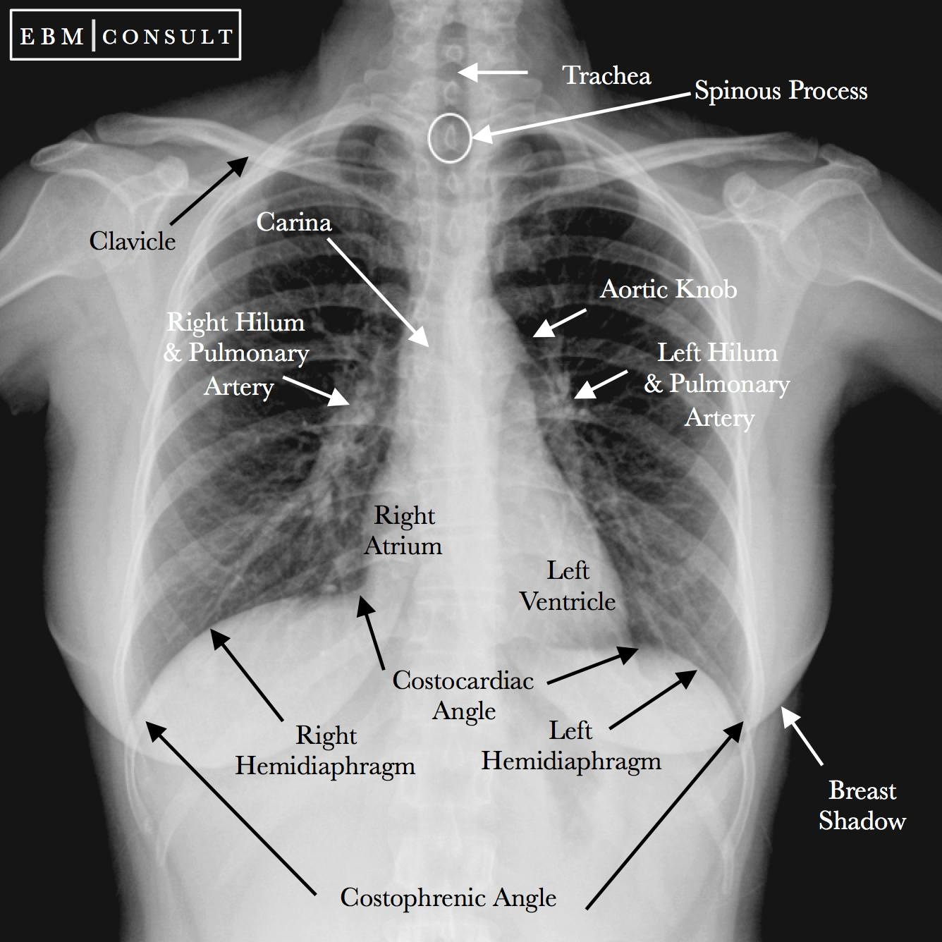
Normal Chest XRay • LITFL Medical Blog • Labelled Radiology

Radiograph of chest in posteroanterior projection. This chapter reacquaints you with the normal anatomy and helps you develop a search pattern that you can apply to every radiograph. There is a degree of hyperinflation as.
Normal Chest XRay • LITFL Medical Blog • Labelled Radiology

Drainage with intercostal (ic) drain. The lungs and pleural spaces are clear. Web ap portable view of the chest; Recent chest infection, spiking temperature. Domes of diaphragm are normal in position and contour.
Normal Chest X Ray Images

The cardiac outlines are normal. Sometimes an exam covers an area of the body but does not discuss any findings. Domes of diaphragm are normal in position and contour. Trachea, carina, bronchi and hilar structures..
Normal chest Xray Stock Image C019/7404 Science Photo Library

High dose antibiotics iv according to cultures/sensitivity. No obvious skeletal abnormality is seen. Follow up of known disease to assess progress. Regularly reviewing and presenting radiographs to senior colleagues while on the ward will help..
Neat How To Report Normal Chest X Ray Write A Good Introduction For
Web the standard radiographic views for evaluation of the chest are the posteroanterior (pa) and lateral projections with the patient standing; High dose antibiotics iv according to cultures/sensitivity. The cardiomediastinal contour is within normal limits..
This study aimed to establish the service enablers and challenges associated with training and. Being familiar with normal anatomy in chest radiographs increases your chances of detecting an abnormality when one is present, even if you can’t diagnose the condition definitively. Trachea, carina, bronchi and hilar structures. Recent chest infection, spiking temperature. Mode of transport of the patient, e.g.
In This Article We Will Focus On:
Being familiar with normal anatomy in chest radiographs increases your chances of detecting an abnormality when one is present, even if you can’t diagnose the condition definitively. Drainage with intercostal (ic) drain. Pa and lateral views of the chest; The anatomy of the chest radiograph ( young adult female ) is well illustrated.
21 Public Playlists Include This Case.
The cardiomediastinal contour is within normal limits. Radiographie du rachis by charles bélanger This section lists what the radiologist saw in each area of the body in the exam. Mode of transport of the patient, e.g.
Radiograph Of A Normal Chest In The A, Posteroanterior And B, Left Lateral Projection.
This is a normal radiograph. Web at a glance. Reading like the pros | radiology key. The cardiac outlines are normal.
Web How To Read A Normal Chest X Ray:
Finalized by the predictable outcome of management, e.g. Sometimes an exam covers an area of the body but does not discuss any findings. Follow up of known disease to assess progress. The lungs and pleural spaces are clear.
Follow up of known disease to assess progress. The lungs and pleural spaces are clear. 21 public playlists include this case. Regularly reviewing and presenting radiographs to senior colleagues while on the ward will help. Pa and lateral views of the chest;